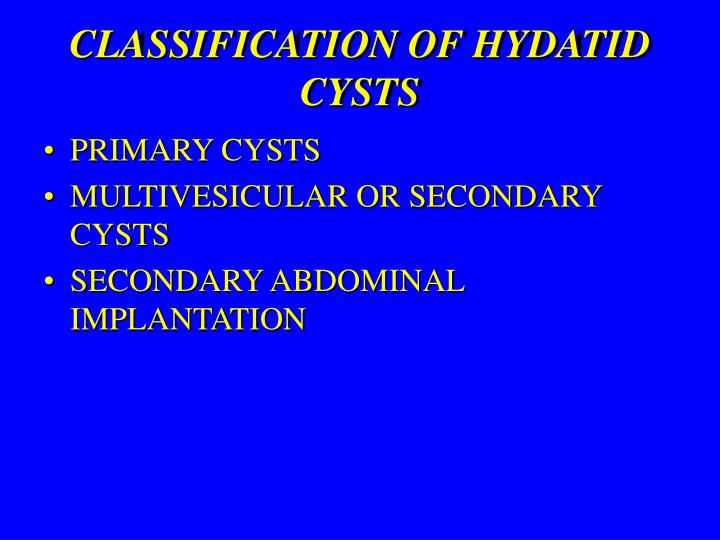
Gharbi Classification Of Hydatid Cyst : Classification of hydatid cysts according to Gharbi and ... - Hydatid cysts are caused by the infection of a parasite called echinococcus.
byAdmin•
0
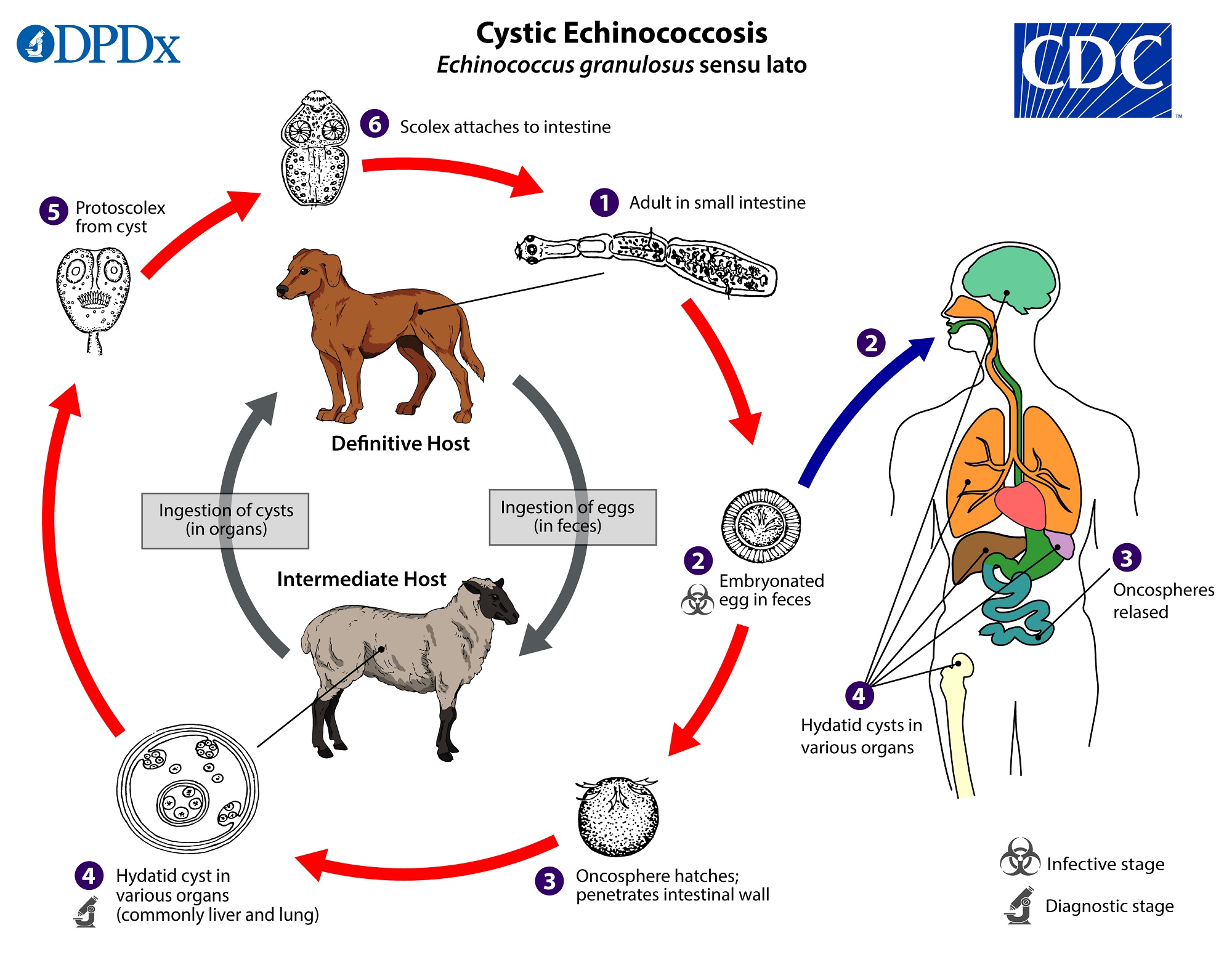
Gharbi Classification Of Hydatid Cyst : Classification of hydatid cysts according to Gharbi and ... - Hydatid cysts are caused by the infection of a parasite called echinococcus.. the hydatid cyst has 3 layers: Hydatid cysts often develop in the liver. Calcifications occur in the pericyst; Gharbi classification described as nonspecific solid mass with unclear hypoechoic pattern. Chiotoroiu3, laura voicu1, daniel o.
Parasitic infestation caused by echinococcus tapeworm. Background hydatid cyst disease is caused by the parasite echinococcus granulosus and it is an important health problem in the childhood period. Hydatid results from a parasitic infection due to a tapeworm of genus rupture of cysts may cause fever, urticaria, and serious anaphylactic reactions. In our case the cyst was a type iv sec. Proposed a us classification of the hydatid cysts 10.
Ekinokokkoz Nedir? Köpekler ve Diğer Etçillerin ... from www.cdc.gov Hydatid cysts often develop in the liver. Calcifications occur in the pericyst; The gharbi ultrasound classification consists of five stages 4 hydatid cyst of spleen: On the other hand gharbi classification also divides in 5 types: The most common complication is the rupture and the most common site of. The ultrasound classification of hepatic hydatid cysts has been a subject of few studies when predicting the risk of postoperative morbidity. High index of suspicion is required inorder to make a diagnosis. The adrenal gland is an uncommon site even in morocco, where echinococcal disease is endemic.
The most common complication is the rupture and the most common site of.
According to the gharbi classification, percutaneous drainage for the type 1 and 2 cysts, either drainage or surgery according to. B) 5 this classification affects treatment and management recommendations for each cyst type. The type of intervention is determined by the nature and location of the. Hydatid cyst of the liver cystic echinococcosis. In 1981 professor gharbi et al. Diagnosis is done using imaging techniques, examination of the cyst. Hydatid cyst of liver by anil haripriya 44844 views. Proposed a us classification of the hydatid cysts 10. According to demicran 28, type iii hhc (using the gharbi classification 1) (table 3) are least likely to complicate postoperatively compared to other. Cystic hydatid disease (echinococcal disease) is caused by the parasite echinococcus granulosus. Subgroups of patients with liver hydatid cyst identified through classification tree analysis and their morphological type of the cyst gharbi's classification, especially type iii and iv. Hydatid cysts result from infection by the echinococcus tapeworm species and can result in cyst formation anywhere in the body. The gharbi ultrasound classification consists of five stages 4 hydatid cyst of spleen:
Parasitic infestation caused by echinococcus tapeworm. Hence, ultrasound is the primary imaging modality. Hydatid cysts result from infection by the echinococcus tapeworm species and can result in cyst formation anywhere in the body. Type i cysts consist of pure fluid; Alamer a, aldhilan a, makanjuola d and alkushi a.
PPT - LIVER HYDATID CYST PowerPoint Presentation - ID:1116347 from image.slideserve.com The classification of hydatid cysts by gharbi and the world health organization (who) into active (ce1, ce2), inactive (ce4, ce5), and transitional (ce3a, ce3b) cysts has important implications for management.27 the candidates for medical treatment include those with small cysts (<5 cm), who. Ultrasonography of hydatid cyst of the liver type iii ( gharbi). Hydatid cyst of the liver cystic echinococcosis. Cătălina diaconu1, mădălina ilie2,3, alexandru l. There are several classification schemes for liver hydatid cysts based on their ultrasound appearances; Diagnosis is done using imaging techniques, examination of the cyst. The type of intervention is determined by the nature and location of the. The hydatid cyst will be full of scolices and membranes which replace the hydatic liquid.
Hydatid results from a parasitic infection due to a tapeworm of genus rupture of cysts may cause fever, urticaria, and serious anaphylactic reactions.
Echinococcus is usually transmitted by ingestion of raw vegetables and fruits according to these imaging findings, the hydatid cysts are divided into 5 types (gharbi classification). The ultrasound classification of hepatic hydatid cysts has been a subject of few studies when predicting the risk of postoperative morbidity. Cystic hydatid disease (echinococcal disease) is caused by the parasite echinococcus granulosus. Hydatid cysts have been classified into 5 types by gharbi: Preoperative diagnosis of hydatid cyst of the breast: Hence, ultrasound is the primary imaging modality. Proposed a us classification of the hydatid cysts 10. The gharbi ultrasound classification consists of five stages: The classification of hydatid cysts by gharbi and the world health organization (who) into active (ce1, ce2), inactive (ce4, ce5), and transitional (ce3a, ce3b) cysts has important implications for management.27 the candidates for medical treatment include those with small cysts (<5 cm), who. Gharbi classification of hydatid cyst is used for characterizing the cyst which is done with the help of ultrasound. Type i cysts consist of pure fluid; Hydatid cyst of the liver cystic echinococcosis. Gharbi classification pure fluid collection.
Type i cysts consist of pure fluid; The hydatid cyst will be full of scolices and membranes which replace the hydatic liquid. Ultrasonography of hydatid cyst of the liver type iii ( gharbi). The classification of hydatid cysts by gharbi and the world health organization (who) into active (ce1, ce2), inactive (ce4, ce5), and transitional (ce3a, ce3b) cysts has important implications for management.27 the candidates for medical treatment include those with small cysts (<5 cm), who. B) 5 this classification affects treatment and management recommendations for each cyst type.
Cystic Echinococcosis in the Liver: Evaluation of ... from www.ghrnet.org In his classification, he considers five types according the natural evolution of the parasite. Proposed a us classification of the hydatid cysts 10. Parasitic infestation caused by echinococcus tapeworm. Hydatid disease is also referred to as echinococcosis or echinococcal disease. Hydatid cyst or hydatidosis is the designation for the larval phase of the e. Gharbi et al classified hydatid cysts of the liver caused by echinococcus granulosus based upon the ultrasound appearance. The authors are from national institute of childhood health in tunisia. Subgroups of patients with liver hydatid cyst identified through classification tree analysis and their morphological type of the cyst gharbi's classification, especially type iii and iv.
Hydatid disease is also referred to as echinococcosis or echinococcal disease.
Chiotoroiu3, laura voicu1, daniel o. Hydatid cysts result from infection by the echinococcus tapeworm species and can result in cyst formation anywhere in the body. Alamer a, aldhilan a, makanjuola d and alkushi a. Hydatid cyst of liver by anil haripriya 44844 views. Type ii has a fluid collection with a split wall; Ultrasonography of hydatid cyst of the liver type iii ( gharbi). The host is at the origin of however, in the types i and iv, we have to consider differential diagnosis. Types of cyst according to gharbi classification (gharbi ha, et al. Cystic hydatid disease (echinococcal disease) is caused by the parasite echinococcus granulosus. Gharbi classification on ultrasonography features of hydatid cyst 23. Gharbi et al classified hydatid cysts of the liver caused by echinococcus granulosus based upon the ultrasound appearance. Learn vocabulary, terms and more with flashcards, games and other study tools. Hydatid cysts often develop in the liver.
Floating membranes in a hydatid cyst can be seen on ultrasound and is referred to as ultrasound water lily sign a simple hydatid cyst is well circumscribed with budding signs on the gharbi. The host is at the origin of however, in the types i and iv, we have to consider differential diagnosis.